L'url de cette page a bien été copiée dans votre presse-papier!
Reference
20230104_0002
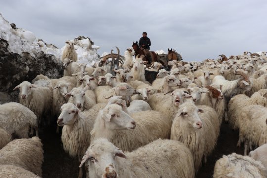
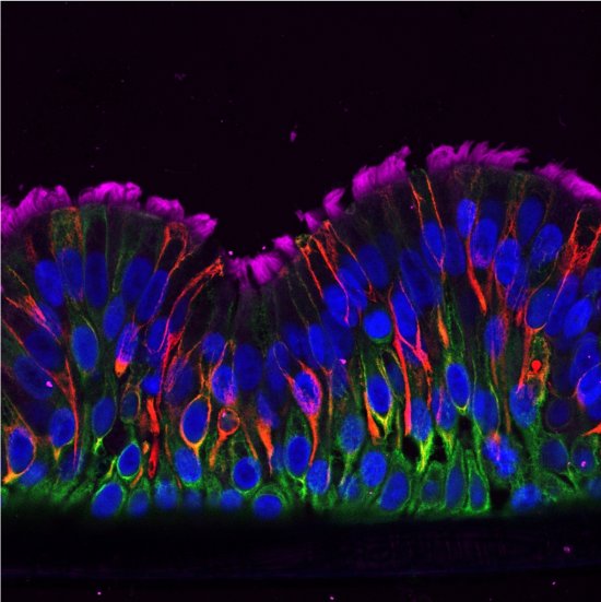
On the tip of the tongue
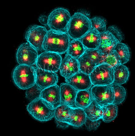
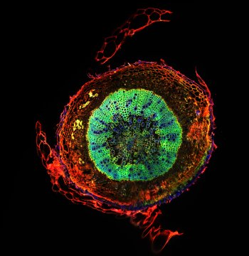
This microscopic image of a sea urchin embryo after five hours of growth reveals the subtle process of cell division. Inside each cell (outlined in turquoise), the microtubules of the mitotic spindle (red) pull the chromosomes (green) in opposite directions, forming two new cells. The sea urchin embryo at an early stage is a biological model as original as it is useful: it helps to identify all the mechanisms that determine cell division orientation in human tissues and the consequences of alterations to these processes, such as the development of certain cancers.
The use of media visible on the CNRS Images Platform can be granted on request. Any reproduction or representation is forbidden without prior authorization from CNRS Images (except for resources under Creative Commons license).
No modification of an image may be made without the prior consent of CNRS Images.
No use of an image for advertising purposes or distribution to a third party may be made without the prior agreement of CNRS Images.
Our work is guided by the way scientists question the world around them and we translate their research into images to help people to understand the world better and to awaken their curiosity and wonderment.